Exam for Group B Streptococcus (Group B Strep or GBS)
What is it?
This is a swab (culture) taken to test for a bacteria known as Group B Strep. Group B Strep is not a sexually transmitted infection (STI)
Why is it done?
- Group B Strep can cause medical problems for your baby after birth.
- If the swab tests positive for Group B Strep or if you have certain risk factors, you’ll be treated with antibiotics during labour. Your baby may also be treated with antibiotics after birth.
How is it done?
At 36 weeks of pregnancy, a swab is taken from your vagina and sometimes from your rectum. This test is usually done at your caregiver’s office and may be done by yourself. It causes no discomfort.
Glucose screening test
What is it?
This is a blood test which may be done at about 24-28 weeks to measure the amount of sugar in your blood.
Why is it done?
- A small number of people develop high blood sugar in pregnancy. This can lead to health problems for your baby before and after birth. It can also affect your health and can cause difficulties during labour and birth.
- It’s usually easy to control high blood sugar. Changes in your diet are often the only treatment required.
How is it done?
A blood sample is taken an hour after you drink a sweet liquid. If your blood sugar is high, further screening will be done.
Ultrasound
What is it?
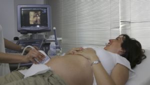
Ultrasound (also called a sonogram) is a test that uses sound echoes to produce images (pictures) of different parts of your body. The scan may take 30 to 60 minutes. You may need to arrange childcare for your older children because children may not be allowed in the ultrasound room. When you are pregnant, an ultrasound can be used to make an image of your baby as it grows and develops in your uterus. The diagnostic centre where you have the ultrasound done will provide instructions.
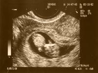
How is it done?
During an ultrasound, sound waves flow into your abdomen through a handheld probe that is gently rolled over your belly. As the sound waves meet structures of different thicknesses within your body, they echo back. These echoes are recorded on a screen similar to a television. The sound waves do not hurt, and because they are beyond the range of human hearing, you will not hear any sound. You may feel pressure on your abdomen as the caregiver moves the probe, but usually no pain. There are no known risks to the parent or baby from this procedure.
Why is it done?
Although ultrasound can be done at any time in pregnancy, the information provided by this test depends on the stage of pregnancy.
Early Viability Scan
In early pregnancy, ultrasound may be used to confirm the gestational age (number of weeks) of the baby, to check if there is more than one baby and to see whether the pregnancy is progressing normally inside the uterus. This scan is useful for people who are having pain or bleeding from the vagina and for people who have had previous miscarriages or ectopic pregnancies.
The 11 to 14 Week Scan or Nuchal Scan
This scan takes place between 11 and 14 weeks and measures the thickness of the layer of fluid at the back of the baby’s neck. The measurement and the results of the maternal blood test are combined with your age:
- to assess the risks of Down Syndrome and other chromosomal abnormalities
- to diagnose certain major fetal abnormalities
- to diagnose early pregnancy failure
The 18 to 20 Week Scan
This is a detailed scan where each part of the baby’s body is checked. Special attention is paid to the brain, face, spine, heart, stomach, bowel, kidneys, arms and legs. If any abnormalities are found, your healthcare provider will be informed and the findings will be discussed with you. Further counselling is available if needed.
The Fetal Well-Being Scan
An ultrasound may be done in the third trimester to check the baby’s growth, follow a suspected or known abnormality, check for complications in high-risk people (for example, those with diabetes or multiple babies), and prepare for birth.
Many caregivers will routinely advise you to have a Fetal Well-Being Scan at about 30 to 32 weeks of pregnancy to check your baby’s growth and well-being. This scan checks the growth and health of the baby using a number of fetal measurements and behaviour observations (the fetal biophysical profile score). The biophysical profile determines your baby’s well-being by measuring the movement of your baby’s body; your baby’s breathing movements, muscle tone of your baby’s arms and legs and the amount of amniotic fluid around your baby. The position and appearance of the placenta and the blood flow to the placenta and baby may be seen by colour Doppler ultrasound. This test takes about 15 minutes to perform.
Maternal Serum Prenatal Screen (MSPS)
What is it?
This is an optional blood test available between 15 and 20 weeks gestation. It measures three substances in the parent’s blood to estimate the chance of having a baby born with chromosomal differences or birth defects: Down Syndrome, Trisomy 18 and neural tube defects (that is, spina bifida, an abnormality of the spine), or birth defects (in the neural tube and abdominal wall). This blood test is also called the “Triple or Quad Screen”
Why is it done?
The MSPS is a screening test, not a diagnostic test. It provides a more accurate estimate of the chance that a pregnancy is affected by either chromosome defects (Down Syndrome or Trisomy 18) or birth defects (in the neural tube or abdominal wall). MSPS is not able to tell with certainty whether a pregnancy is affected with any of these conditions. If your MSPS results indicate that you have an increased chance of having a baby with a concern, follow-up will be offered. This may involve genetic counselling to discuss the results and the option of amniocentesis and/or ultrasound. It is your choice whether or not to have any further prenatal testing. MSPS does not screen for all chromosome problems or health concerns.
How is it done?
A lab requisition can be obtained from your healthcare provider in order to get your blood drawn. The best time to do this test is between 15 and 16 weeks gestation, however the test can be done up to 20 weeks. It cannot be done before 15 weeks, and accurate gestational dating is important. Results are usually available within 2 to 3 working days.
Please contact your caregiver or a genetic counsellor if you have any questions.
Amniocentesis
What is it?
This is a medical procedure performed by a doctor that involves using a needle to remove a small sample of amniotic fluid from around the baby. The amniotic fluid contains cells and proteins made by the baby and the placenta. The fluid is tested in the laboratory.
Why is it done?
Amniocentesis may be done to determine if your baby has a chromosome problem (for example, Down Syndrome). It can also detect the majority of neural tube defects like spina bifida. Later in pregnancy, it may also be done to test the maturity of the baby’s lungs. It is not a routine procedure, but is offered in pregnancies that are at an increased risk for a health problem.
Who is offered an amniocentesis?
It is a standard of care to offer amniocentesis to people who:
- are 35 years of age or older.
- have a screening test that shows an increased risk (screen positive).
- have a family or personal history that puts the pregnancy at increased risk.
How is it done?
Ultrasound is used to determine the position of your baby. A fine needle is carefully inserted through the abdomen and a small amount of amniotic fluid is removed.
What are the risks of amniocentesis?
There are risks to amniocentesis. The most significant is an increased risk of miscarriage above the baseline population rate. In most cases, the increase in risk is about 0.5%, or 1 in every 200 amniocentesis procedures that are performed. It is the patient’s choice whether or not to have this procedure done. Genetic counselling is available to get more information about amniocentesis before making the decision to have this procedure. Please contact your caregiver or a genetic counsellor if you have any questions.
Chorionic Villus Sampling (CVS)
This screening test may not be available in your area.
What is it?
CVS is a test used to get a sample from the placenta between 10 and 14 weeks of pregnancy.
Why is it done?
Since the genetic make-up of the cells of the placenta are usually the same as the baby, this tissue can be used for diagnosing certain problems with the baby. It can diagnose the same conditions as an amniocentesis but can be offered earlier in pregnancy.
How is it done?
The first step of the CVS test is an ultrasound examination to find the placenta, and measure the baby to determine the gestational age of the baby (number of weeks). Ultrasound is then used to guide a thin biopsy forcep through the cervix and into the placenta (transcervical CVS). Another method uses a thin needle inserted through the abdomen into the placenta (transabdominal CVS). A small piece of the placenta is removed and examined under a microscope. Most of the time the sample is enough and it is not necessary to get a second specimen. You may have minor cramping and vaginal bleeding when the transcervical procedure is done. Local anesthesia (freezing) may be used for the transabdominal procedure.
What are the risks with CVS?
- The natural rate of pregnancy loss after 10 weeks is about 5%. After CVS, the risk of miscarriage is increased by about 1%.
- Cramping, spotting, and bleeding can happen after CVS. These symptoms do not usually last long and do not negatively affect the pregnancy.
- Infection after CVS is very rare. If you have fever, abdominal pain or vaginal discharge within 24 to 72 hours, call your healthcare provider.
- Rarely, the healthcare provider will not be able to obtain an adequate sample of chorionic villi. If results are desired, you will be offered a repeat CVS or amniocentesis later in the pregnancy.
What tests are done with CVS tissue?
- Chromosome analysis
- DNA testing: DNA studies may be done when there is a family history of a genetic disorder.
NOTE:
Alpha-fetoprotein (AFP) levels are measured in the parent’s blood or amniotic fluid. This test can help determine if there is an open neural tube defect such as spina bifida. AFP cannot be measured in CVS tissue. Before having CVS you should have counselling to help you understand the risks, benefits and limitations of the procedure. Early prenatal diagnosis is the main advantage of CVS compared to amniocentesis.
Fetal Monitoring
What is it?
Fetal monitoring is done to assess the well-being of your baby at every prenatal check up, it is also done during a non-stress test and/or a biophysical profile . The non-stress test is a type of electronic fetal monitoring which measures how your baby’s heart rate reacts to his or her own body movement.
Why is it done?
Electronic fetal monitoring records your baby’s heartbeat. It also checks your baby’s well-being after 24 weeks of pregnancy.
How is it done?
During check ups a hand held monitor is held against your abdomen to listen to the baby’s heartbeat. During the non-stress test two sensors are placed on your abdomen that are held lightly in place by elastic belts. One sensor measures your baby’s heart rate and the other sensor measures contractions. The test takes about 20 minutes.
Counting Baby Movements
What is it?
Healthcare providers sometimes suggest using a Fetal Movement Chart to track your baby’s movements more accurately after 30 weeks.
Why is it done?
- Counting movements each day is a simple way to check your baby’s health.
- An active baby is usually a healthy baby.
- Decrease in movement may be the first warning that your baby is not well or is having trouble.
- A lack of or decrease in movement does not occur before labour starts. Even when you’re in labour, you should still feel your baby move.
How is it done?
Fetal Movement Count sheets and instructions on how to use them are available through your healthcare provider.